This is my personal story of having tummy tuck surgery (abdominoplasty) with lateral thigh lift, including before & after pictures and a daily log of my pre-operation, day-of-surgery and post-operation recovery experiences. If you are a man looking to get one of these, this is what you may be in for.

Male Tummy Tuck With Body Lift
A tummy tuck (also known as Abdominoplasty) is elective cosmetic surgery of the abdomen for removing excess skin and fat (saggy skin and wrinkles) and to tighten the skin over the stomach. It is the only known remedy to remove baggy skin that is so stretched out that that it has lost it’s elasticity (test your skin elasticity) and no amount of exercise or proper nutrition can restore it.
I am a 54 year old man who had attained a weight of over 300 pounds. I am 5′-7″ tall. My maximum weight was probably over 300 pounds, but I really don’t know because I stopped looking at the scale. I do remember being weighed at the doctor’s office and my weight was 298 lbs., but I’m sure I gained more weight after that.
Over the course of about 2 or 3 years of dieting and daily exercise, I reduced my weight to 155 lbs. (I lost over 150 pounds). This left me with a lot of excess skin around my belly and midsection. Since I didn’t want to live with this for the rest of my life, I decided to look into surgery.
The first thing I wondered was “Have I lost enough weight to have an operation?” It was difficult for me to tell. Was my enlarged midsection (see my before photos) still due to fat or due to stretched skin. I researched ideal weight charts for men, BMIs, average weights and more in an effort to determine if I was ready. I decided that I was and thought that if I wasn’t, the doctor would tell me so. In hindsight, I probably should have lost 5-10 more pounds. I did ask the doctor if I should lose some more weight and he would not say. He didn’t indicate why. I think he was afraid that if he did say that I was ready and had the operation with results that I was not satisfied with, I would blame him. I now think it best to go all the way down to 5 lbs. or more below your ideal target weight just to be on the safe side. That way, after the operation the stomach would be nice and flat.
Do Men Have Tummy Tucks?
I looked at all the before and after pictures that I could find and read all the books, news articles, blogs and information on the internet concerning this type of plastic surgery for men (male tummy tucks). It turns out that there is much more information about this subject that is geared towards women than there is about tummy tucks for men. Just about all of the tummy tuck forums and discussion boards that I visited were almost exclusively for women. In 2008, only 3% of the surgeries performed in the United States were for men. I think this may change in the future with the increasing obesity problems we now face. Also there appears to be a stigma or shame associated with tummy tucks for men. I know I was initially embarrassed when I first made inquires regarding my operation.
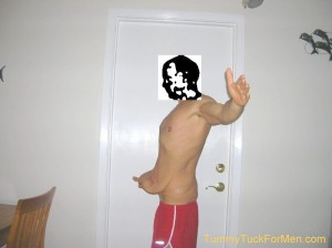
Excess Skin Before Surgery

Day After Surgery
I also read about the horror stories and the success stories. Those horror stories as well as all the “bad stuff” that can happen to you on the operating table and during recovery really makes you think twice as to whether it’s worth it. Then I looked into all of the alternatives but found they couldn’t fix my problem. After looking in the mirror a few more times and doing some soul searching, I decided to risk it and looked into finding a plastic surgeon in my area. I was fortunate to find an excellent surgeon in my city. On 9/17 I called to make an appointment for a consultation.
Tummy Tuck For Men Surgery
To remove excess skin from the abdominal area the surgeon first makes a long incision from hip to hip just above the pubic area. He then makes a second incision around the navel (the navel is the scar on your abdomen that was caused when the umbilical cord was removed when you were a baby. It can be either an innie (depression) or an outie (protrusion). The shape and appearance of the navel is a result of the scar, looseness of the skin, fat under the skin, surrounding musculature and the abdominal wall. Little known fact - since navels are scars and are not a makeup of genetics, they can be used to distinguish between identical twins) to free the belly button from the surrounding skin. The skin is then surgically separated from the abdominal wall up to the ribs and lifted to expose the muscles in the abdomen. The muscles are repositioned and stitched to provide a firmer, slimmer and more contoured abdomen. The skin is then stretched back into place and a new hole is cut for the belly button which is then stitched in place. Unwanted excess skin is cut away and the incision is closed with stitches.
(9/24) Going to his office for the initial visit was sort of intimidating, being a 54 year old male who hasn’t been to many doctors, especially not a plastic surgeon with a waiting room full of women. Fortunately, both he and his staff were friendly and made the visit easier.
My thoughts on my drive to my first consultation included:
- Will I be naked for the “Before” Photo?
- The Horror Stories that you read about.
- Is this guy really a good plastic surgeon?
- Will I need liposuction as well?
- Have I lost enough weight?
- This is not going to be covered by insurance.
- “Tummy tuck” is a foolish name to say out loud and abdominoplasty is too hard to remember.
First Tummy Tuck Surgery Consultation
I was lead into an examining room where a nurse provided me with additional forms and literature and answered most of the tummy tuck questions that I had prepared. The initial examination by the doctor fortunately only involved stripping down to my underwear (being there was humiliating enough). It was a “brief” examination by the doctor who indicated that a tummy tuck as well as a Lateral Thigh Lift would be appropriate for me, but that the decision was entirely mine. He indicated that I did not need liposuction and that he could not do it in conjunction with this type of operation anyway because of the complexity of it. I was shown before and after photos of previous tummy tucks that the doctor had performed and received a more complete description of the operation and potential complications. The nurse provided me with written price quotes for the procedures and a schedule and asked if I wanted to proceed with the operation. I told her I would consider it and call back for another appointment. I was charged a $25 for my consultation fee and was quoted the following prices. I was surprised to find that they required payment in full prior to the operation. They also required a $500 deposit when they scheduled the operation. Good thing they took Amex.
Price Quote for Male Tummy Tuck Surgery with Lateral Thigh Lift: $11,200
The cost Included doctor’s fee, facility and support personnel – $10,000 for the doctor’s fee and $1200 for the anesthesia fee.
Price Quote for Medial Thigh Lift: $6,350
$5,500 for the doctors fee and $850 for the anesthesia fee.
Types Of Tummy Tuck Plastic Surgery
- Tummy tuck surgery is where they cut away and stitch “tight” the sagging skin in be belly area. The medical term for “tummy tuck” is Abdominoplasty. It is a surgical procedure involving the removal of excess skin and fat from the middle and lower parts of the abdomen (stomach, belly, tummy) and sometimes tightening the abdomen muscles resulting in a flatter abdomen. Scars associated with the surgery are usually hidden below the line of a swimsuit or underwear. Many scars fade with time but do not go away completely.
- Outer thigh lift (lateral) surgery is where they remove the sagging skin from the outer thighs and may require operating on the buttocks area. In my case that meant continuing along the sides of the waist and back area and do the same thing to lift the hips and the butt.
- Inner thigh lift (medial) surgery is where they remove the sagging skin from the inside area of the thighs. Operating on this area can remove sagging skin, improve the overall appearance and reduce the rubbing together of the inner thighs. Liposuction may be used to remove excess fat. I decided against having the Medial Thigh Lift performed because for me, it would have to be a separate operation, cost more money and I figured I could live with what I had.
- Lower body lift (belt lipectomy) surgery removes sagging skin from the tummy, thighs and the buttocks.
My Next Surgery Consultation
A few weeks later I made another appointment where I received additional paperwork and information as how to proceed. They also gave me a “Medical Clearance for Surgery” form for me to get signed by my family doctor. That meant another trip to my doctor for an additional examination including blood testing, urine analysis and an EKG test. Usually chest x-rays are performed, but since I was a non-smoker, that was not required.
Since I would need continual care and monitoring for 24 hours after the surgery, I needed some additional help. They provided me with the name and number of a home healthcare nurse. I called and made arrangements with Donna (the nurse) to pick me up, take me to surgery, take me home and stay with me overnight tending to my needs.
Getting My Family Doctor’s Approval for Surgery (10/3)
I went to my family doctor for the required examination and tests and to get the “Medical Clearance for Surgery” form signed by the doctor. I needed blood tests and an EKG (electrocardiogram is a recording of the electrical activity of the heart) for him to complete the form.
Back to the Surgeon’s Office (11/9)
I paid the $500 deposit and scheduled the surgery date for December 4.
Last consultation Before Surgery (11/20)
I provided the signed Medical Clearance for Surgery form (medical clearance) to the receptionist and paid the balance in full ($10,700) with my Amex credit card. The doctor took the “Before” photographs. I told him I didn’t want to see these plastered all over the internet (I was holding a sign with my name on it). He said not to worry. I was then provided surgery pre-operation and post-operation instructions and I received prescriptions to get filled prior to the day of the operation (12/4/08). The two prescriptions that I received were for Keflex 500mg an antibiotic (20 pills) and Mepergan Fortis for pain (40 pills).
I Was All Set For My Tummy Tuck Surgery
Now all I had to do was wait and follow the Surgery Pre-Operation and Post-Operation Instructions and my Preparation for Surgery Checklist that I created.
See what happened on the Day Of My Tummy Tuck Surgery.
About Tummy Tuck For Men
Tummy Tuck For Men is a web site specifically dedicated to providing information about abdominoplasty plastic surgery. It is created and updated by an individual with regular entries of personal commentary, descriptions of events, pictures, video or other material. A blog is usually displayed in reverse-chronological order and is a form of an online diary.
Why I wrote Tummy Tuck For Men
I am a 54 year old man who is 5′-7 1/2″ tall who had attained a weight of over 300 pounds. After over 2 years of dieting and daily exercise I reduced my weight to 155lbs. This left me with a lot of excess skin around my belly and mid section. Since I didn’t want to live with this for the rest of my life, I decided to look into an operation.
I never thought that I would ever write a blog. Getting a tummy tuck (especially for a guy) is a very personal matter. Publicly writing about it would be impossible for me to do (or so I thought).
I decided to document each detail of my experience on my computer for my personal records. I also took lots of “before & after” photographs because I wanted to see for myself the changes it would make.
The pictures and information stayed in my computer for over a year. Then I realized that I had regained 15 pounds (it just sort of snuck up on me). I could not get heavy again.
I needed some incentive to keep the weight off. Hopefully writing about it would give me that incentive. So I changed my mind about making my experience public. Self-motivation was my primary reason. I had gained 15 lbs over the year and needed to take it off before it got out of control. In an effort to keep me focused on a lifestyle of health, exercise and good nutrition I decided to create a blog or website describing my experience of having the surgery and include all the information that I would have wanted to see when I first started doing my initial research a year ago.
Note - After my blog became established on the internet and started to receive traffic I added advertising to try to make a little extra money. This was not my main reason for publishing Tummy Tuck For Men but an after-thought. By writing and regularly maintaining this blog, I remain focused on health, nutrition and exercise. This has provided the required motivation to be fit, healthy and maintain my ideal weight. But, It is still a work in progress.
Leave a Reply